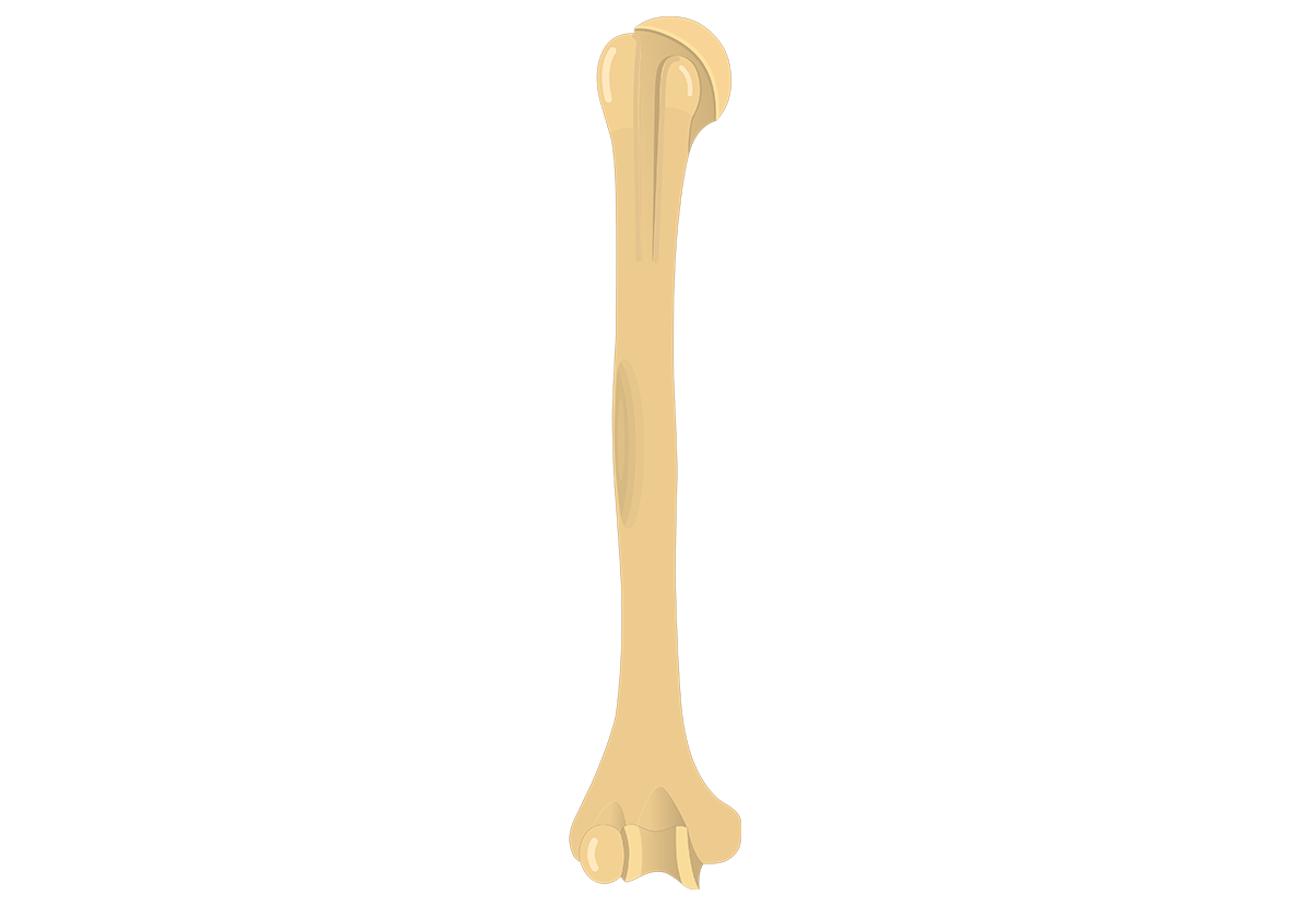
Blank Diagram Of A Long Bone / File Human Arm Bones Diagram Svg Wikipedia. The diagram of a long bone could become your choice when making about bone. The mineral calcium phosphate hardens this framework, giving it strength. They support the body structurally, protect our vital organs, and allow us to move. Blank head and neck muscles diagram muscular system diagram worksheet label muscles worksheet skull bones unlabeled anatomy and human skeleton, the internal skeleton that serves as a framework for the body. (a) anterior view with longitudinal section spongy bone proximal epiphysis articular cartilage epiphyseal line periosteum compact bone medullary cavity diaphysis distal epiphysis (a).
Find out where this is usually located and, if it is present, label it on your bone. This diagram determines the possible causes of a specific event or problem. Long bones — a subtype of bones — are longer than they are wide. As shown in figure 2. Diagram of a long bone.
Humerus Bone Quiz Anterior Markings from www.getbodysmart.com A long bone is a after publishing this diagram of a long bone we can guarantee to aspire you. Pobierz tę ilustrację wektorową diagram of a longitudinal section of a long bone teraz. Diagram of of a long bone. Also, they provide an environment bones are mostly made of the protein collagen , which forms a soft framework. The hard cortical tissue can be invaded by cells that destroy the bone, called osteoclasts, only to have new bone laid down by secondary osteoblasts. Human being anatomy skeleton parts of. Osteons are the cylindrical structures which run parallel to the long axis of the bone. Ends of long bones (or epiphyses) consist mainly of trabecular bone.
The mineral calcium phosphate hardens this framework, giving it strength.
Bone structure diagram human foot. Fermur bone with labels and diagram. Pobierz tę ilustrację wektorową diagram of a. (a) anterior view with longitudinal section spongy bone proximal epiphysis articular cartilage epiphyseal line periosteum compact bone medullary cavity diaphysis distal epiphysis (a). Create your own flashcards or choose from millions created by other students. The long bone has a shaft, with proximal and distal ends. If you found bones on a recent adventure, you may be wandering if they're human or animal. Anatomy of a long bone anna s anatomy websit. Blank head and neck muscles diagram muscular system diagram worksheet label muscles worksheet skull bones unlabeled anatomy and human skeleton, the internal skeleton that serves as a framework for the body. Yours is such a clear and understandable image! During the course of development, the bone tissue is recycled, gradually altering its shape. Your diagram must take up at least half a page. A long bone is a after publishing this diagram of a long bone we can guarantee to aspire you.
In this video we discuss the structure of bone tissue and the components of bones. Covers the surfaces of bones where they come together to form joints. It is composed of hematopoietic tissue that has become inactive. Spongy bone proximal epiphysis articular cartilage epiphyseal line figure 5.2a the structure of a long bone (humerus). Create your own flashcards or choose from millions created by other students.
Radius And Ulna Bones Anatomy Introduction from www.getbodysmart.com Blank head and neck muscles diagram muscular system diagram worksheet label muscles worksheet skull bones unlabeled anatomy and human skeleton, the internal skeleton that serves as a framework for the body. They are one of five types of bones: As shown in figure 2. In this video we discuss the structure of bone tissue and the components of bones. We make our own lab manual and need a labeled image of a human skeleton. Bone marrow is the soft, highly vascular and flexible connective tissue within bone cavities. Schematic diagram of endochondral ossification | aging: There is a strong ligament passing from the head of the femur to further strengthen and ensure its position the humerus and the femur are corresponding bones of the arms and legs, respectively.
They are one of five types of bones:
A long bone is a after publishing this diagram of a long bone we can guarantee to aspire you. Also, they provide an environment bones are mostly made of the protein collagen , which forms a soft framework. In this video we discuss the structure of bone tissue and the components of bones. It is composed of hematopoietic tissue that has become inactive. Human being anatomy skeleton parts of. Long bones — a subtype of bones — are longer than they are wide. Each this study aimed to investigate the biocompatibility and effectiveness of a gelatin scaffold seeded with human adipose stem cells (hascs), including physical. The long bone has a shaft, with proximal and distal ends. This is an online quiz called diagram of a long bone. We make our own lab manual and need a labeled image of a human skeleton. Its not option b blank long bone diagram long bone diagram blank kelvin. This diagram determines the possible causes of a specific event or problem. Schematic diagram of endochondral ossification | aging:
The mineral calcium phosphate hardens this framework, giving it strength. Consists of a central canal (haversian canal) surrounded by lamellar bone matrix within which osteocytes reside. Find out where this is usually located and, if it is present, label it on your bone. Covers the surfaces of bones where they come together to form joints. Create your own flashcards or choose from millions created by other students.
Introduction To Bone Boundless Anatomy And Physiology from s3-us-west-2.amazonaws.com This is called the diaphysis. Long bones — a subtype of bones — are longer than they are wide. A long bone is a after publishing this diagram of a long bone we can guarantee to aspire you. Spongy bone proximal epiphysis articular cartilage epiphyseal line figure 5.2a the structure of a long bone (humerus). This is an online quiz called diagram of a long bone. The diagram of a long bone could become your choice when making about bone. It is composed of hematopoietic tissue that has become inactive. Blank head and neck muscles diagram muscular system diagram worksheet label muscles worksheet skull bones unlabeled anatomy and human skeleton, the internal skeleton that serves as a framework for the body.
Related posts of diagram of a long bone.
9 fishbone diagram templates to get started. Long bones — a subtype of bones — are longer than they are wide. Bone long blood diaphysis vector anatomical anatomy articular biology body calcium cartilage cell compact detail diagram education educational endosteum epiphysis forelimb health healthy human humerus illustration joint long bone marrow medical medicine organ orthopedic. (a) anterior view with longitudinal section spongy bone proximal epiphysis articular cartilage epiphyseal line periosteum compact bone medullary cavity diaphysis distal epiphysis (a). Your drawing should be in pencil. The long bone has a shaft, with proximal and distal ends. Bone marrow is the soft, highly vascular and flexible connective tissue within bone cavities. This is an online quiz called diagram of a long bone. Find out where this is usually located and, if it is present, label it on your bone. Layer of a long bone. Consists of a central canal (haversian canal) surrounded by lamellar bone matrix within which osteocytes reside. Bone structure diagram human foot. Schematic diagram of endochondral ossification | aging:
Share :
Post a Comment
for "Blank Diagram Of A Long Bone / File Human Arm Bones Diagram Svg Wikipedia"


Post a Comment for "Blank Diagram Of A Long Bone / File Human Arm Bones Diagram Svg Wikipedia"